- Date:2020-01-08
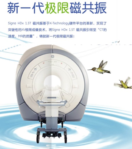
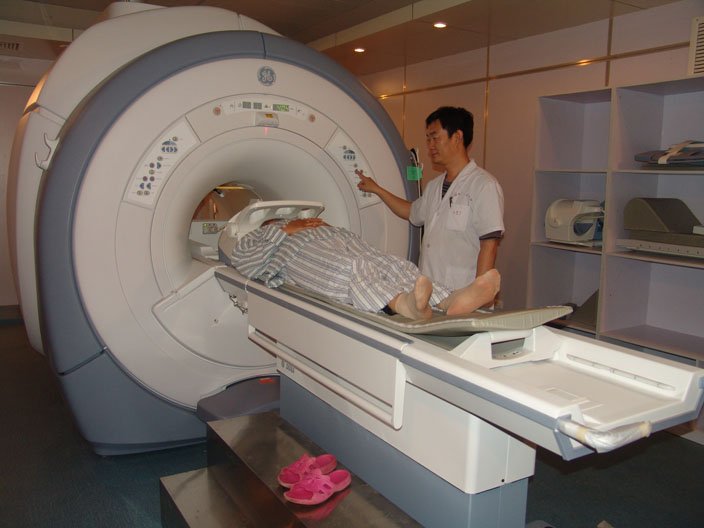
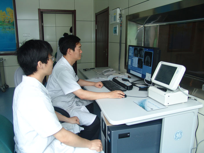
MR room was established in March 1998, the first in the city to introduce Japan Hitachi low-field magnetic resonance, after more than ten years of development, so that the hospital's imaging technology has been in the city's leading level. 2010 June and the introduction of the U.S. GE high configuration 1.5T superconducting magnetic resonance scanning machine (SIGNA HDx), will be a powerful impetus to promote diagnostic level of Lucy's imaging, and to better serve the people. Service. There are 7 people in the MR room of our hospital, and the director of the department, Pang Shanjun, has 1 deputy chief physician, 2 attending physicians, 3 physicians, and 1 technician. They have studied in the General Hospital of the People's Liberation Army, Tiantan Hospital, the First Affiliated Hospital of Xi'an Jiaotong University, the Third Hospital of North Medicine and other teaching hospitals, which is a ladder-type professional scientific and technological team. The department has published more than 20 papers in national and provincial magazines, completed 3 scientific and technological achievements independently and with the participation of the department, and won the second and third prizes for scientific and technological progress in Liaocheng City. There are 2 three patents and 3 books.
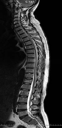
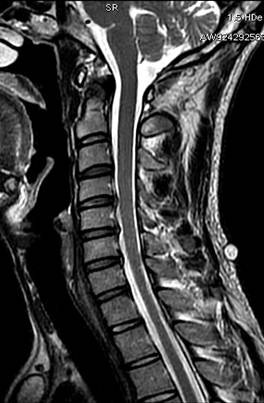
The department carries out the following services: in addition to the routine examination of all parts of the body, the department carries out head and neck joint enhanced angiography, chest and abdomen enhanced angiography, lower limb three-segment enhanced angiography, cerebral white matter fibre bundle imaging, cranial cerebral perfusion imaging (DSC and ASL-PWI), magnetic susceptibility imaging (SWAN), single-voxel and multiple-voxel spectral imaging; pancreatico-cholangiography, urinary imaging, otorhinolaryngology, and other imaging techniques. urography, cochlear hydrography, whole spine scanning, whole body diffusion imaging (PET-like), etc.; and the introduction of breast and cardiac 8-channel phased array coils, which are able to fully meet the needs of patients and clinics for breast and cardiac function tests.
Our Signa 1.5T HDx MRI machine is GE's 5th generation magnetic resonance imaging system, which adopts XV technology, which is regarded as one of the greatest revolutionary technologies of the 21st century and the main direction of the future development of magnetic resonance systems, and its hardware advantages are mainly reflected in:
§ Limit imaging XV technology platform, fully optimised RF system, to achieve the effect of zero transmission, zero reconstruction of the revolutionary breakthrough. Under the condition of 256*256 matrix and uncompressed data, the reconstruction speed of post-processing per second reaches the industry's highest 2700 frames per second, which really achieves the speed of CT and the quality of MR!
§ Three-dimensional volumetric self-calibrating spatial parallel acquisition technology, completely eliminating misalignment artefacts, shortening the time of LAVA-XV (full abdominal volumetric perfusion imaging) to 9 seconds (the original 17 seconds), and expanding the scope to 90 layers of the whole abdomen, with a layer thickness of 2mm (the original can only be done for part of the liver, with a layer thickness of 3-4mm).
§ HDx's exclusive ECO Extreme Gradient System turbocharges the industry with the shortest TR time and shortest TE time. The shortest TR time determines the image scanning speed, the shorter the faster; the shortest TE time determines the image quality, the shorter the higher signal-to-noise ratio.
Its clinical advantages are mainly reflected in:
§ LAVA-XV - 9-second full abdominal high-resolution perfusion imaging
§ Propeller-XV - successful examination of uncooperative children in a single session without sedation
§ Cardiac-XV cardiac acquisition quality has reached a new height, cardiac Cardiac-xv unique local uniform field technology, completely overcome the artifacts of the stubborn disease, automatic shortest TR/TE time real-time completion of myocardial survival examination, paediatric congenital heart disease and myocardial disease examination is more accurate, more rapid
GE's high-quality magnets provide the industry's best magnetic field uniformity, providing a guarantee of high-quality imaging images.
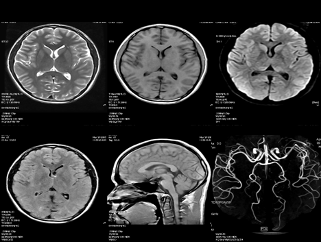
Liver Plain Scanning and 3D Volumetric Dynamic Enhancement Scanning

Two-dimensional and three-dimensional magnetic resonance cholangiopancreatography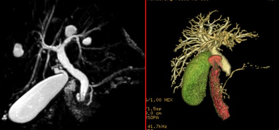
Abdominal Magnetic Resonance Angiography Comparable to DSA

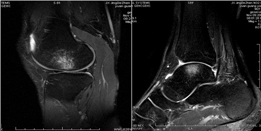
Weighted image of joint fat suppression
Scanning for dirty hearts

Coronal and sagittal images of the pelvis
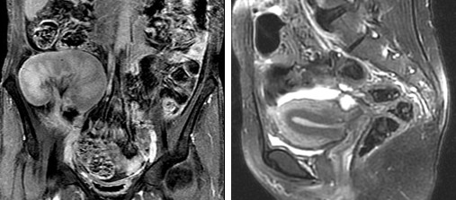
Indications of MRI examination
1、Cranial brain and spinal cord MRI is more sensitive than CT in the diagnosis of brain tumour, encephalitis, cerebral white matter lesions, cerebral infarction, congenital brain anomalies, etc., and can detect early lesions and locate them more accurately. MRI can directly display some cranial nerves, and can find early lesions occurring on these nerves.MRI can directly display the whole picture of spinal cord, so it has important diagnostic value for spinal cord tumour or intravertebral canal tumour, spinal cord leukoencephalopathy, spinal cord cavitation, spinal cord injury and so on. MRI can directly show the whole spinal cord. For intervertebral disc lesions, MRI can show its degeneration, herniation or bulging. It also shows spinal stenosis better. For cervical and thoracic spine, CT often shows unsatisfactory, while MRI shows clearly. In addition, MRI is also very sensitive to show metastatic tumours in the vertebral body.
Head and neck MRI is good for displaying tumourous lesions in eye, ear, nose, throat, such as nasopharyngeal cancer invasion of skull base and cranial nerves, which is clearer and more accurate than CT.MRI can also do angiography of neck and show vascular anomalies. MRI can also show the range and characteristics of neck lumps to help characterisation.
Chest MRI can directly display the myocardium and the left and right ventricular chambers (gated with electricity), which can be used to understand the damage to the myocardium and determine the function of the heart. The condition of large vessels in the mediastinum can be clearly shown. It is also extremely helpful in the localisation and characterisation of mediastinal tumours. It can also show pulmonary oedema, pulmonary embolism and lung tumours. It can distinguish the nature of pleural effusion, vascular section or lymph node.
Abdominal MRI can provide valuable information for the diagnosis of liver, kidney, pancreas, spleen, adrenal gland and other substantial organs, which can help to confirm the diagnosis. It is also easier to show small lesions, and thus can detect early lesions.MR cholangiopancreatography (MRCP) can show the bile duct and pancreatic duct, which can replace ERCP.MR urography (MRU) can show the dilated ureter and renal pelvis and kidney calyces, which is especially suitable for patients with poor renal function, and IVU does not show the image.
5、Pelvis MRI can show lesions of uterus, ovary, bladder, prostate, seminal vesicles and other organs. It can directly see the endometrium and myometrium, which is very helpful for early diagnosis of tumourous lesions in the uterus. It is also of great value for the localisation and qualitative diagnosis of lesions in ovary, bladder and prostate.
Posterior peritoneum MRI is of great value in showing the tumour of the posterior peritoneum and its relationship with the surrounding organs. It can also show the lesions of abdominal aorta or other large vessels, such as abdominal aortic aneurysm, Buchanan syndrome, renal artery stenosis and so on.
7, musculoskeletal system MRI on the cartilage discs in the joints, tendons, ligaments, the display rate is higher than CT. As it is more sensitive to the changes of bone marrow, it can find bone metastasis, osteomyelitis, aseptic necrosis, leukaemia bone marrow infiltration at an early stage. It shows clearly on soft tissue mass of bone tumour. It also has certain diagnostic value for soft tissue injury.
Precautions for MRI examination
(a) What should be done to prepare the patient before MRI examination?
1. Before entering the examination room, patients must take out all metal objects on their bodies, such as mobile phones, watches, keys, pens, coins, glasses, and various magnetic cards.
2, for young children, restlessness and depression and phobia patients to give the right amount of sedatives.
3、Abdominal examination is best done on an empty stomach, and gastrointestinal contrast can be taken.
(ii) Which patients should not undergo MRI scanning?
1、People with cardiac pacemakers.
2. Those with arterial clips after aneurysm surgery.
(3) Those who have metal foreign bodies in the eyeball.
4. Critically ill patients with all kinds of resuscitation equipment.
5, patients with various metal implants in the body should be cautious when checking.
6, agitation can not be suppressed.
(C) MRI examination has contraindications?
Yes. Contraindication is that the patient's body is equipped with magnetic susceptibility of substances or devices, the movement of these structures or loss of function will cause adverse consequences. Such as:
1. cardiac pacemakers;
2. cochlear implants;
3. certain artificial heart valves;
4. bone growth stimulators and nerve stimulators (TENs);
5. arterial clips or coils;
6. metal structures;
7. certain prostheses.
Liaocheng Second People's Hospital
Magnetic resonance room
Appointment Consultation Tel: 0635-2342341, 2342681



 鲁ICP备11009722号-4